The Ultimate Curiosity

Brainstorming is our aim.

The Ultimate Curiosity

Brainstorming is our aim.

Heart Bypass Surgery Explained with Video

Before your surgery you will get general anesthesia. You will be asleep (unconscious) and pain-free during surgery. Once you are unconscious, the heart surgeon will make a 8-10-inch surgical cut (incision) in the middle ...

The Ultimate Curiosity

Brainstorming is our aim.

The Ultimate Curiosity

Brainstorming is our aim.

Sunday 3 June 2012

Protozoan Diseases of the Cardiovascular and Lymphatic Systems


Trypanosoma cruzi causes Chagas’ disease. The reservoir includes many wild animals. The vector is reduviid, the “kissing bug.”
Xenodiagnosis allows for the identification of trypanosomes in the intestinal tract of the reduviid bug, which confirms the diagnosis. 


                                                                       Trypanosoma cruzi 


Toxoplasmosis

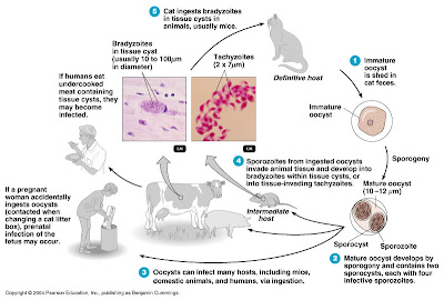
Toxoplasmosis is caused by the sporozoan Toxoplasma gondii.
T. gondii undergoes sexual reproduction in the intestinal tract of domestic cats (the reservoir), and oocysts are eliminated in cat feces.
In the host cell, sporozoites reproduce to form either tissue invading tachyzoites or bradyzoites.
Humans contract the infection by ingesting tachyzoites or tissue cysts in undercooked meat from an infected animal or contact with cat feces (transmission is gastrointestinal).
Congenital infections can occur. Signs and symptoms include severe brain damage or vision problems.
Toxoplasmosis can be identified by serological tests, but interpretation of the results is uncertain. 


Malaria

The signs and symptoms of malaria are chills, fever, vomiting, and headache, which occur at intervals of 2-3 days.
Malaria is transmitted by Anopheles mosquitoes. The causative agent is any one of four species of Plasmodium.
Sporozoites reproduced in the liver and release merozoites into the blood stream, where they infect red blood cells and produce more merozoites.
Laboratory diagnosis is based on microscopic observation of merozoites in red blood cells.
New drugs are being developed as the protozoa develop resistance to drugs such as chloroquine.
Malaria in the United States
                         

Malaria


Leishmaniasis

Leishmania spp., which are transmitted by sandflies, cause leishmaniasis.
Leishmania donovani - visceral leishmaniasis: the protozoa reproduce in the liver, spleen and kidneys.
Leishmania tropica - cutaneous leishmaniasis (Oriental sore): affects skin
Leishmania braziliensis - mucocutaneous leishmaniasis: affects mucous membranes as well as skin
Antimony compounds are used for treatment.
Cutaneous Leishmaniasis

Babesiosis

Babesiosis is caused by the protozoan Babesia microti and transmitted to humans by ticks.

Helminthic Diseases of the Cardiovascular and Lymphatic Systems

Schistosomiasis

Species of the blood fluke Schistosoma cause schistosomiasis.
Eggs eliminated with feces hatch into larvae that infect the intermediate host, a snail. Free-swimming cercariae are released from the snail and penetrate the skin of a human.
The adult flukes live in the veins of the liver or urinary bladder in humans.
Granulomas are from the host’s defense against eggs that remain in the body.
Observation of eggs or flukes in feces, skin tests, or indirect serological tests may be used for diagnosis.
Chemotherapy (praziquantel or oxamniquine) is used to treat the disease; sanitation and snail eradication are used to prevent it.
Schistosomiasis
Schistosome Granuloma

Swimmer’s Itch

Swimmer’s itch is a cutaneous allergic reaction to cercariae that penetrate the skin. The definitive hosts for this fluke are wildfowl.


Friday 18 May 2012

ATP: The Perfect Energy Currency for the Cell


Abstract

The major energy currency molecule of the cell, ATP, is evaluated in the context of creationism. This complex molecule is critical for all life from the simplest to the most complex. It is only one of millions of enormously intricate nanomachines that needs to have been designed in order for life to exist on earth. This motor is an excellent example of irreducible complexity because it is necessary in its entirety in order for even the simplest form of life to survive.

Introduction

IIn order to function, every machine requires specific parts such as screws, springs, cams, gears, and pulleys. Likewise, all biological machines must have many well-engineered parts to work. Examples include units called organs such as the liver, kidney, and heart. These complex life units are made from still smaller parts called cells which in turn are constructed from yet smaller machines known asorganelles. Cell organelles include mitochondria, Golgi complexes, microtubules, and centrioles. Even below this level are other parts so small that they are formally classified as macromolecules (large molecules).
Fig. 1. Views of ATP and related structures.
A critically important macromolecule—arguably “second in importance only to DNA”—is ATP. ATP is a complexnanomachine that serves as the primary energy currency of the cell (Trefil, 1992, p.93). A nanomachine is a complex precision microscopic-sized machine that fits the standard definition of a machine. ATP is the “most widely distributed high-energy compound within the human body” (Ritter, 1996, p. 301). This ubiquitous molecule is “used to build complex molecules, contract muscles, generate electricity in nerves, and light fireflies. All fuel sources of Nature, all foodstuffs of living things, produce ATP, which in turn powers virtually every activity of the cell and organism. Imagine the metabolic confusion if this were not so: Each of the diverse foodstuffs would generate different energy currencies and each of the great variety of cellular functions would have to trade in its unique currency” (Kornberg, 1989, p. 62).
ATP is an abbreviation for adenosine triphosphate, a complex molecule that contains the nucleoside adenosine and a tail consisting of three phosphates. (See Figure 1 for a simple structural formula and a space filled model of ATP.) As far as known, all organisms from the simplest bacteria to humans use ATP as their primary energy currency. The energy level it carries is just the right amount for most biological reactions. Nutrients contain energy in low-energy covalent bonds which are not very useful to do most of kinds of work in the cells.
These low energy bonds must be translated to high energy bonds, and this is a role of ATP. A steady supply of ATP is so critical that a poison which attacks any of the proteins used in ATP production kills the organism in minutes. Certain cyanide compounds, for example, are poisonous because they bind to the copper atom in cytochrome oxidase. This binding blocks the electron transport system in the mitochondria where ATP manufacture occurs (Goodsell, 1996, p.74).  

The Aufbau Principle


The electron configuration of an atom gives the distribution  of electrons among atomic orbitals in the atom.  Two general methods are used to show electron configurations.  The subshell notation uses numbers to designate the principal shells and the letters s, p, d, and f to identify the subshells.  A superscript following the letter indicates the number of electrons in the designated subshell.  The ground state electron configuration for nitrogen would be   1s22s22p3.  A drawback to this method of showing the electron configuration is that it does not tell us how the three 2p electrons are distributed among the three 2p orbitals.  We can show this by using an orbital diagram in which boxes are used to indicate orbitals within subshells and arrows to represent electrons in these orbitals.  The direction of the arrows represent the directions of the electron spins.  The orbital diagram for nitrogen is
1s
2s
 
2p
 
The way we arrive at electron configurations such as the one for nitrogen above is to use a set of rules collectively called the aufbau principle. 
  • Electrons occupy orbitals of the lowest energy available
  • No two electrons in the same atom may have all four quantum numbers alike
  • When entering orbitals of the same energy, electrons initially occupy them singly ant with the same spin
  • Electrons fill orbitals in order of the quantum number sum (n + l). For equal (n + l) sums, fill levels in order of increasing n.
A mnemonic diagram for the aufbau principle known as the diagonal rule is shown here
The aufbau principle is really a thought process in which we think about building up an atom from the one that preceeds it in atomic number, by adding a proton and neutrons to the nucleus and one electron to the appropriate atomic orbital.  
There are some exceptions to the to the aufbau principle.  The first is chromium (Z = 24), the aufbau principle predicts the an electron configuration of  [Ar]3d44s2 but experimentally we find it to be  [Ar]3d54s1.  The next exception found is that of copper (Z = 29), the predicted electron configuration is  [Ar]3d94s2 but experimentally we find it to be  [Ar]3d104s1.  The reason for these and other exceptions are not completely understood, but it seems that a half-filled 3d subshell in the case of chromium or a completely-filled  3d subshell in the case of copper lend a special stabilty to the observed electron configurations.  There is no need to dwell on these exceptions, the point to remember is that the aufbau principle predicts the correct electron configuration most of the time and that the energy of the predicted electron configuration is very close to the ground state energy.

Thursday 17 May 2012

Artificial respiration


Artificial respiration is the act of assisting or stimulating respiration, a metabolic process referring to the overall exchange of gases in the body by pulmonary ventilation, external respiration, and internal respiration. Assistance takes many forms, but generally entails providing air for a person who is not breathing or is not making sufficient respiratory effort on their own(although it must be used on a patient with a beating heart or as part of cardiopulmonary resuscitation to achieve the internal respiration).
Pulmonary anton ventilation (and hence external parts of respiration) is achieved through manual insufflation of the lungs either by the rescuer blowing into the patient's lungs, or by using a mechanical device to do so. This method of insufflation has been proved more effective than methods which involve mechanical manipulation of the patient's chest or arms, such as the Silvester method. It is also known as Expired Air Resuscitation (EAR), Expired Air Ventilation (EAV), mouth-to-mouth resuscitationrescue breathing or colloquially the kiss of life.
Artificial respiration is a part of most protocols for performing cardiopulmonary resuscitation(CPR) making it an essential skill for first aid. In some situations, artificial respiration is also performed separately, for instance in near-drowning and opiate overdoses. The performance of artificial respiration in its own is now limited in most protocols to health professionals, whereas lay first aiders are advised to undertake full CPR in any case where the patient is not breathing sufficiently.
Mechanical ventilation involves the use of a mechanical ventilator to move air in and out of the lungs when an individual is unable to breathe on his or her own, for example during surgery withgeneral anesthesia or when an individual is in a coma.

Insufflations


Insufflation, also known as 'rescue breaths' or 'ventilations', is the act of mechanically forcing air into a patient's respiratory system. This can be achieved via a number of methods, which will depend on the situation and equipment available. All methods require good airway management to perform, which ensures that the method is effective. These methods include:
  • Mouth to mouth - This involves the rescuer making a seal between their mouth and the patient's mouth and 'blowing', to pass air into the patient's body
  • Mouth to nose - In some instances, the rescuer may need or wish to form a seal with the patient's nose. Typical reasons for this include maxillofacial injuries, performing the procedure in water or the remains of vomit in the mouth
  • Mouth to mouth and nose - Used on infants (usually up to around 1 year old), as this forms the most effective seal
  • Mouth to mask – Most organisations recommend the use of some sort of barrier between rescuer and patient to reduce cross infection risk. One popular type is the 'pocket mask'. This may be able to provide higher tidal volumes than a Bag Valve Mask.
  • Bag valve mask (BVM) - This is a simple device manually operated by the rescuer, which involves squeezing a bag to expel air into the patient.
  • Mechanical resuscitator - An electric unit designed to breathe for the patient.

Adjuncts to insufflation

.
A CPR pocket mask, with carrying case
Most training organisations recommend that in any of the methods involving mouth to patient, that a protective barrier is used, to minimise the possibility of cross infection (in either direction).
Barriers available include pocket masks and keyring-sized face shields. These barriers are an example of Personal Protective Equipment to guard the face against splashing, spraying or splattering of blood or other potentially infectious materials.
These barriers should provide a one-way filter valve which lets the air from the rescuer deliver to the patient while any substances from the patient (e.g. vomit, blood) cannot reach the rescuer. Many adjuncts are single use, though if they are multi use, after use of the adjunct, the mask must be cleaned and autoclaved and the filter replaced.
The CPR mask is the preferred method of ventilating a patient when only one rescuer is available. Many feature 18mm inlets to support supplemental oxygen, which increases the oxygen being delivered from the approximate 17% available in the expired air of the rescuer to around 40-50%.
Tracheal intubation is often used for short term mechanical ventilation. A tube is inserted through the nose (nasotracheal intubation) or mouth (orotracheal intubation) and advanced into the trachea. In most cases tubes with inflatable cuffs are used for protection against leakage and aspiration. Intubation with a cuffed tube is thought to provide the best protection against aspiration. Tracheal tubes inevitably cause pain and coughing. Therefore, unless a patient is unconscious or anesthetized for other reasons, sedative drugs are usually given to provide tolerance of the tube. Other disadvantages of tracheal intubation include damage to the mucosal lining of the nasopharynx or oropharynx and subglottic stenosis.
In an emergency a Cricothyrotomy can be used by health care professionals, where an airway is inserted through a surgical opening in the cricothyroid membrane. This is similar to atracheostomy but a cricothyrotomy is reserved for emergency access. This is usually only used when there is a complete blockage of the pharynx or there is massive maxillofacial injury, preventing other adjunts being used .

Efficiency of mouth to patient insufflation

Normal atmospheric air contains approximately 21% oxygen when created in. After gaseous exchange has taken place in the lungs, with waste products (notably carbon dioxide) moved from the bloodstream to the lungs, the air being exhaled by humans normally contains around 17% oxygen. This means that the human body utilises only around 19% of the oxygen inhaled, leaving over 80% of the oxygen available in the exhalatory breath.
This means that there is more than enough residual oxygen to be used in the lungs of the patient, which then crosses the cell membrane to form oxyhemoglobin.

Oxygen 


The efficiency of artificial respiration can be greatly increased by the simultaneous use of oxygen therapy. The amount of oxygen available to the patient in mouth to mouth is around 16%. If this is done through a pocket mask with an oxygen flow, this increases to 40% oxygen. If a Bag Valve Mask or mechanical respirator is used with an oxygen supply, this rises to 99% oxygen. The greater the oxygen concentration, the more efficient the gaseous exchange will be in the lungs.


Wednesday 16 May 2012

Bone grafting


Bone grafting is a surgical procedure that replaces missing bone in order to repair bone fractures that are extremely complex, pose a significant health risk to the patient, or fail to heal properly.
Bone generally has the ability to regenerate completely but requires a very small fracture space or some sort of scaffold to do so. Bone grafts may be autologous (bone harvested from the patient’s own body, often from the iliac crest), allograft (cadaveric bone usually obtained from a bone bank), or synthetic (often made ofhydroxyapatite or other naturally occurring and biocompatible substances) with similar mechanical properties to bone. Most bone grafts are expected to be reabsorbed and replaced as the natural bone heals over a few months’ time.
The principles involved in successful bone grafts include osteoconduction (guiding the reparative growth of the natural bone), osteoinduction (encouraging undifferentiated cells to become active osteoblasts), and osteogenesis (living bone cells in the graft material contribute to bone remodeling). Osteogenesis only occurs with autografts.

Biological mechanism

Properties of various types of bone graft sources.
OsteoconductiveOsteoinductiveOsteogenic
Alloplast+
Xenograft+
Allograft++/–
Autograft+++
Bone grafting is possible because bone tissue, unlike most other tissues, has the ability to regenerate completely if provided the space into which to grow. As native bone grows, it will generally replace the graft material completely, resulting in a fully integrated region of new bone. The biologic mechanisms that provide a rationale for bone grafting are osteoconduction, osteoinduction and osteogenesis

Osteoconduction

Osteoconduction occurs when the bone graft material serves as a scaffold for new bone growth that is perpetuated by the native bone. Osteoblasts from the margin of the defect that is being grafted utilize the bone graft material as a framework upon which to spread and generate new bone. In the very least, a bone graft material should be osteoconductive.

Osteoinduction

Osteoinduction involves the stimulation of osteoprogenitor cells to differentiate into osteoblasts that then begin new bone formation. The most widely studied type of osteoinductive cell mediators are bone morphogenetic proteins (BMPs). A bone graft material that is osteoconductive and osteoinductive will not only serve as a scaffold for currently existing osteoblasts but will also trigger the formation of new osteoblasts, theoretically promoting faster integration of the graft.


Osteopromotion

Osteopromotion involves the enhancement of osteoinduction without the possession of osteoinductive properties. For example,enamel matrix derivative has been shown to enhance the osteoinductive effect of demineralized freeze dried bone allograft(DFDBA), but will not stimulate de novo bone growth alone.


Osteogenesis

Osteogenesis occurs when vital osteoblasts originating from the bone graft material contribute to new bone growth along with bone growth generated via the other two mechanisms

Types and Tissue Sources


Autograft

Autologous (or autogenous) bone grafting involves utilizing bone obtained from the same individual receiving the graft. Bone can be harvested from non-essential bones, such as from the iliac crest, or more commonly in oral and maxillofacial surgery, from the mandibular symphysis (chin area) or anterior mandibular ramus (the coronoid process); this is particularly true for block grafts, in which a small block of bone is placed whole in the area being grafted. When a block graft will be performed, autogenous bone is the most preferred because there is less risk of the graft rejection because the graft originated from the patient's own body. As indicated in the chart above, such a graft would be osteoinductive and osteogenic, as well as osteoconductive. A negative aspect of autologous grafts is that an additional surgical site is required, in effect adding another potential location for post-operative pain and complications.
Autologous bone is typically harvested from intra-oral sources as the chin or extra-oral sources as the iliac crest, the fibula, the ribs, the mandible and even parts of the skull.
All bone requires a blood supply in the transplanted site. Depending on where the transplant site is and the size of the graft, an additional blood supply may be required. For these types of grafts, extraction of the part of the periosteum and accompanying blood vesels along with donor bone is required. This kind of graft is known as a vital bone graft.
An autograft may also be performed without a solid bony structure, for example using bone reamed from the anterior superior iliac spine. In this case there is an osteoinductive and osteogenic action, however there is no osteoconductive action, as there is no solid bony structure.

Allografts

Allograft bone, like autogenous bone, is derived from humans; the difference is that allograft is harvested from an individual other than the one receiving the graft. Allograft bone is taken from cadavers that have donated their bone so that it can be used for living people who are in need of it; it is typically sourced from a bone bank.
There are three types of bone allograft available:
  1. Fresh or fresh-frozen bone
  2. Freeze-dried bone allograft (FDBA)
  3. Demineralized freeze-dried bone allograft (DFDBA)

Synthetic variants

Artificial bone can be created from ceramics such as calcium phosphates (e.g. hydroxyapatite and tricalcium phosphate), Bioglass and calcium sulphate; all of which are biologically active to different degrees depending on solubility in the physiological environment. These materials can be doped with growth factors, ions such as strontium or mixed with bone marrow aspirate to increase biological activity. Some authors believe this method is inferior to autogenous bone grafting however infection and rejection of the graft is much less of a risk, the mechanical properties such as Young's modulus are comparable to bone. The presence of elements such as strontium can result in higher bone mineral density and enhanced osteoblast proliferation in vivo

Xenografts

Xenograft bone substitute has its origin from a species other than human, such as bovine. Xenografts are usually only distributed as a calcified matrix. In January 2010 Italian scientists announced a breakthrough in the use of wood as a bone substitute, though this technique is not expected to be used for humans until at the earliest 2015.


Alloplastic grafts

Alloplastic grafts may be made from hydroxylapatite, a naturally occurring mineral that is also the main mineral component of bone. They may be made from bioactive glass. Hydroxylapetite is a Synthetic Bone Graft, which is the most used now among other synthetic due to its osteoconduction, hardness and acceptability by bone. Some synthetic bone grafts are made of calcium carbonate, which start to decrease in usage because it is completely resorbable in short time which make the bone easy to break again. Finally used is the tricalcium phosphate which now used in combination with hydroxylapatite thus give both effect osteoconduction and resorbability.


Growth Factors

Growth Factor enhanced grafts are produced using recombinant DNA technology. They consist of either Human Growth Factors or Morphogens (Bone Morphogenic Proteins in conjunction with a carrier medium, such as collagen).


Low HDL Cholesterol (Hypoalphalipoproteinemia)


Low levels of high-density lipoprotein cholesterol (HDL), or hypoalphalipoproteinemia (HA), includes a variety of conditions, ranging from mild to severe, in which concentrations of alpha lipoproteins or high-density lipoprotein (HDL) are reduced. The etiology of HDL deficiencies ranges from secondary causes, such as smoking, to specific genetic mutations, such as Tangier disease and fish-eye disease.
HA has no clear-cut definition. An arbitrary cutoff is the 10th percentile of HDL cholesterol levels. A more practical definition derives from the theoretical cardioprotective role of HDL. The US National Cholesterol Education Program (NCEP) Adult Treatment Panel III (ATP III) redefined the HDL cholesterol level that constitutes a formal coronary heart disease (CHD) risk factor. The level was raised from 35 mg/dL to 40 mg/dL for men and women. A prospective analysis by Mora et al investigated the link between cholesterol and cardiovascular events in women and found baseline HDL-C level was consistently and inversely associated with incident coronary and CVD events across a range of LDL-C values.
For the metabolic syndrome in which multiple mild abnormalities in lipids, waist size (abdominal circumference), blood pressure, and blood sugar increase the risk of CHD, the designated HDL cholesterol levels that contribute to the syndrome are sex-specific. For men, a high-risk HDL cholesterol level is still less than 40 mg/dL, but for women, the high-risk HDL cholesterol level is less than 50 mg/dL.
A low HDL cholesterol level is thought to accelerate the development ofatherosclerosis because of impaired reverse cholesterol transport and possibly because of the absence of other protective effects of HDL, such as decreased oxidation of other lipoproteins.
The common, mild forms of HA have no characteristic physical findings, but patients may have premature coronary heart or peripheral vascular disease, as well as a family history of low HDL cholesterol levels and premature CHD.
Therapy to raise the concentration of HDL cholesterol includes weight loss, smoking cessation, aerobic exercise, and pharmacologic management with niacin and fibrates.
This review addresses the pathogenesis and presenting features of, and the diagnostic tests, therapeutic interventions, and follow-up strategies for, low HDL cholesterol levels.