The Ultimate Curiosity

Brainstorming is our aim.

The Ultimate Curiosity

Brainstorming is our aim.

Heart Bypass Surgery Explained with Video

Before your surgery you will get general anesthesia. You will be asleep (unconscious) and pain-free during surgery. Once you are unconscious, the heart surgeon will make a 8-10-inch surgical cut (incision) in the middle ...

The Ultimate Curiosity

Brainstorming is our aim.

The Ultimate Curiosity

Brainstorming is our aim.

Showing posts with label medical science. Show all posts

Sunday, 3 June 2012

Protozoan Diseases of the Cardiovascular and Lymphatic Systems


Trypanosoma cruzi causes Chagas’ disease. The reservoir includes many wild animals. The vector is reduviid, the “kissing bug.”
Xenodiagnosis allows for the identification of trypanosomes in the intestinal tract of the reduviid bug, which confirms the diagnosis. 


                                                                       Trypanosoma cruzi 


Toxoplasmosis

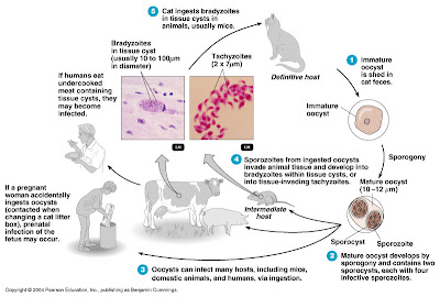
Toxoplasmosis is caused by the sporozoan Toxoplasma gondii.
T. gondii undergoes sexual reproduction in the intestinal tract of domestic cats (the reservoir), and oocysts are eliminated in cat feces.
In the host cell, sporozoites reproduce to form either tissue invading tachyzoites or bradyzoites.
Humans contract the infection by ingesting tachyzoites or tissue cysts in undercooked meat from an infected animal or contact with cat feces (transmission is gastrointestinal).
Congenital infections can occur. Signs and symptoms include severe brain damage or vision problems.
Toxoplasmosis can be identified by serological tests, but interpretation of the results is uncertain. 


Malaria

The signs and symptoms of malaria are chills, fever, vomiting, and headache, which occur at intervals of 2-3 days.
Malaria is transmitted by Anopheles mosquitoes. The causative agent is any one of four species of Plasmodium.
Sporozoites reproduced in the liver and release merozoites into the blood stream, where they infect red blood cells and produce more merozoites.
Laboratory diagnosis is based on microscopic observation of merozoites in red blood cells.
New drugs are being developed as the protozoa develop resistance to drugs such as chloroquine.
Malaria in the United States
                         

Malaria


Leishmaniasis

Leishmania spp., which are transmitted by sandflies, cause leishmaniasis.
Leishmania donovani - visceral leishmaniasis: the protozoa reproduce in the liver, spleen and kidneys.
Leishmania tropica - cutaneous leishmaniasis (Oriental sore): affects skin
Leishmania braziliensis - mucocutaneous leishmaniasis: affects mucous membranes as well as skin
Antimony compounds are used for treatment.
Cutaneous Leishmaniasis

Babesiosis

Babesiosis is caused by the protozoan Babesia microti and transmitted to humans by ticks.

Helminthic Diseases of the Cardiovascular and Lymphatic Systems

Schistosomiasis

Species of the blood fluke Schistosoma cause schistosomiasis.
Eggs eliminated with feces hatch into larvae that infect the intermediate host, a snail. Free-swimming cercariae are released from the snail and penetrate the skin of a human.
The adult flukes live in the veins of the liver or urinary bladder in humans.
Granulomas are from the host’s defense against eggs that remain in the body.
Observation of eggs or flukes in feces, skin tests, or indirect serological tests may be used for diagnosis.
Chemotherapy (praziquantel or oxamniquine) is used to treat the disease; sanitation and snail eradication are used to prevent it.
Schistosomiasis
Schistosome Granuloma

Swimmer’s Itch

Swimmer’s itch is a cutaneous allergic reaction to cercariae that penetrate the skin. The definitive hosts for this fluke are wildfowl.


Thursday, 29 March 2012

Nuclear medicine


Nuclear medicine is a medical specialty involving the application of radioactive substances in the diagnosis and treatment of disease.
In nuclear medicine procedures, radionuclides are combined with other elements to form chemical compounds, or else combined with existing pharmaceutical compounds, to form radiopharmaceuticals. These radiopharmaceuticals, once administered to the patient, can localize to specific organs or cellular receptors. This property of radiopharmaceuticals allows nuclear medicine the ability to image the extent of a disease-process in the body, based on the cellular function and physiology, rather than relying on physical changes in the tissue anatomy. In some diseases nuclear medicine studies can identify medical problems at an earlier stage than other diagnostic tests. It would not be wrong to call Nuclear Medicine as "Radiology done inside out" or "Endo-radiology" because it records radiation emitting from within the body rather than radiation that is generated by external sources like Xrays.
Treatment of diseased tissue, based on metabolism or uptake or binding of a particular ligand, may also be accomplished, similar to other areas of pharmacology. However, the treatment effects of radiopharmaceuticals rely on the tissue-destructive power of short-range ionizing radiation.
In the future, nuclear medicine may provide added impetus to the field known as molecular medicine. As our understanding of biological processes in the cells of living organism expands, specific probes can be developed to allow visualization, characterization, and quantification of biologic processes at the cellular and subcellular levels.[1]Nuclear medicine is an ideal specialty to adapt to the new discipline of molecular medicine, because of its emphasis on function and its utilization of imaging agents that are

Diagnostic medical imaging

Diagnostic

In nuclear medicine imaging, radiopharmaceuticals are taken internally, for example intravenously or orally. Then, external detectors (gamma cameras) capture and form images from the radiation emitted by the radiopharmaceuticals. This process is unlike a diagnostic X-ray where external radiation is passed through the body to form an image.
There are several techniques of diagnostic nuclear medicine.
  • 2D: Scintigraphy ("scint") is the use of internal radionuclides to create two-dimensional[2] images. '
  • 3D: SPECT is a 3D tomographic technique that uses gamma camera data from many projections and can be reconstructed in different planes. Positron emission tomography (PET) uses coincidence detection to image functional processes.
Nuclear medicine tests differ from most other imaging modalities in that diagnostic tests primarily show the physiological function of the system being investigated as opposed to traditional anatomical imaging such as CT or MRI. Nuclear medicine imaging studies are generally more organ or tissue specific (e.g.: lungs scan, heart scan, bone scan, brain scan, etc.) than those in conventional radiology imaging, which focus on a particular section of the body (e.g.: chest X-ray, abdomen/pelvis CT scan, head CT scan, etc.). In addition, there are nuclear medicine studies that allow imaging of the whole body based on certain cellular receptors or functions. Examples are whole body PET scan or PET/CT scans, gallium scans, indium white blood cell scans, MIBG and octreotide scans.
Iodine-123 whole body scan for thyroid cancer evaluation. The study above was performed after the total thyroidectomy and TSH stimulation with thyroid hormone medication withdrawal. The study shows a small residual thyroid tissue in the neck and a mediastinum lesion, consistent with the thyroid cancer metastatic disease. The uptakes in the stomach and bowel are normal physiologic findings.
While the ability of nuclear metabolism to image disease processes from differences in metabolism is unsurpassed, it is not unique. Certain techniques such as fMRI image tissues (particularly cerebral tissues) by blood flow, and thus show metabolism. Also, contrast-enhancement techniques in both CT and MRI show regions of tissue which are handling pharmaceuticals differently, due to an inflammatory process.
Diagnostic tests in nuclear medicine exploit the way that the body handles substances differently when there is disease or pathology present. The radionuclide introduced into the body is often chemically bound to a complex that acts characteristically within the body; this is commonly known as a tracer. In the presence of disease, a tracer will often be distributed around the body and/or processed differently. For example, the ligand methylene-diphosphonate (MDP) can be preferentially taken up by bone. By chemically attachingtechnetium-99m to MDP, radioactivity can be transported and attached to bone via the hydroxyapatite for imaging. Any increased physiological function, such as due to a fracture in the bone, will usually mean increased concentration of the tracer. This often results in the appearance of a 'hot-spot' which is a focal increase in radio-accumulation, or a general increase in radio-accumulation throughout the physiological system. Some disease processes result in the exclusion of a tracer, resulting in the appearance of a 'cold-spot'. Many tracer complexes have been developed to image or treat many different organs, glands, and physiological processes.

Hybrid scanning techniques

In some centers, the nuclear medicine scans can be superimposed, using software or hybrid cameras, on images from modalities such as CT or MRI to highlight the part of the body in which the radiopharmaceutical is concentrated. This practice is often referred to as image fusion or co-registration, for example SPECT/CT and PET/CT. The fusion imaging technique in nuclear medicine provides information about the anatomy and function, which would otherwise be unavailable, or would require a more invasive procedure or surgery.

Wednesday, 28 March 2012

Echocardiography


An echocardiogram, often referred to in the medical community as a cardiac ECHO or simply an ECHO, is a sonogram of the heart. (It is not abbreviated as ECG, which in medicine usually refers to an electrocardiogram.) Also known as a cardiac ultrasound, it uses standard ultrasound techniques to image two-dimensional slices of the heart. The latest ultrasound systems now employ 3D real-time imaging.
In addition to creating two-dimensional pictures of the cardiovascular system, an echocardiogram can also produce accurate assessment of the velocity of blood and cardiac tissue at any arbitrary point using pulsed or continuous wave Doppler ultrasound. This allows assessment of cardiac valve areas and function, any abnormal communications between the left and right side of the heart, any leaking of blood through the valves (valvular regurgitation), and calculation of the cardiac output as well as the ejection fraction. Other parameters measured include cardiac dimensions (luminal diameters and septal thicknesses) and E/A ratio.
Echocardiography was an early medical application of ultrasound. Echocardiography was also the first application of intravenouscontrast-enhanced ultrasound. This technique injects gas-filled microbubbles into the venous system to improve tissue and blood delineation. Contrast is also currently being evaluated for its effectiveness in evaluating myocardial perfusion. It can also be used with Doppler ultrasound to improve flow-related measurements (see Doppler echocardiography).
Echocardiography is performed by cardiac sonographers, cardiac physiologists (UK) or doctors trained in cardiology.

Three-dimensional echocardiography

3D echocardiography is now possible, using an ultrasound probe with an array of transducers and an appropriate processing system. This enables detailed anatomical assessment of cardiac pathology, particularly valvular defects, and cardiomyopathies. The ability to slice the virtual heart in infinite planes in an anatomically appropriate manner and to reconstruct Three-dimensional images of anatomic structures make 3D echocardiography unique for the understanding of the congenitally malformed heart. Real Time 3-Dimensional echocardiography can be used to guide the location of bioptomes during right ventricular endomyocardial biopsies.
The 3D Echo Box developed by the European Association of Echocardiography offers a complete review of Three Dimensional Echocardiography.


Cardiac catheterization


Cardiac catheterization (heart cath) is the insertion of a catheter into a chamber or vessel of the heart. This is done for both investigational and interventional purposes. Subsets of this technique are mainly coronary catheterization, involving the catheterization of the coronary arteries, and catheterization of cardiac chambers and valves.


Coronary catheterization


Procedure

Local anaesthetic is injected into the skin, usually in the right groin, to numb the area. In some centers access to the coronary arteries is made via the right radial or brachial artery (hand or arm), but the majority of cases are still done from the groin region. A puncture is then made with a needle in either the femoral artery in the groin or the radial artery in the wrist, (Seldinger technique), before a guidewire is inserted into the arterial puncture. A plastic sheath (with a stiffer plastic introducer inside it) is then threaded over the wire and pushed into the artery. The wire is then removed and the side-port of the sheath is aspirated to ensure arterial blood flows back. It is then flushed with saline. This arterial sheath, with a bleedback prevention valve, acts as a conduit into the artery for the duration of the procedure.
Catheters are inserted using a guidewire and moved towards the heart. Once in position above the aortic valve the guidewire is then removed. The catheter is then engaged with the origin of the coronary artery (either left main stem or right coronary artery) and x-ray opaque iodine-based contrast is injected to make the coronary vessels show up on the x-ray fluoroscopy image.
When the necessary procedures are complete, the catheter is removed. Firm pressure is applied to the site to prevent bleeding. This may be done by hand or with a mechanical device. Other closure techniques include an internal suture and plug. If the femoral artery was used, the patient will probably be asked to lie flat for several hours to prevent bleeding or the development of a hematoma. If the arm is used, the patient can ambulate sooner. Cardiac interventions such as the insertion of a stent prolong both the procedure itself as well as the post-catheterization time spent in allowing the wound to clot.
A cardiac catheterization is a general term for a group of procedures that are performed using this method, such as coronary angiography, as well as left ventrical angiography. Once the catheter is in place, it can be used to perform a number of procedures including angioplasty, PCI (percutaneous coronary intervention) angiographyballoon septostomy, and an Electrophysiology study.

Indications for investigational use

This technique has several goals:

Electrocardiography


Electrocardiography (ECG or EKG from the German Elektrokardiogramm) is a transthoracic (across the thorax or chest) interpretation of the electrical activity of the heart over a period of time, as detected by electrodes attached to the outer surface of the skin and recorded by a device external to the body.[1] The recording produced by this noninvasive procedure is termed as electrocardiogram(also ECG or EKG). An electrocardiogram (ECG) is a test that records the electrical activity of the heart.

ECG is used to measure the rate and regularity of heartbeats as well as the size and position of the chambers, the presence of any damage to the heart, and the effects of drugs or devices used to regulate the heart (such as a pacemaker). See also stress test andHolter monitor (24h).
The etymology of the word is derived from the Greek electro, because it is related to electrical activity, kardioGreek for heart, andgraph, a Greek root meaning "to write".
Most ECGs are performed for diagnostic or research purposes on human hearts, but may also be performed on animals, usually for research


Function

The ECG device detects and amplifies the tiny electrical changes on the skin that are caused when the heart muscle depolarizes during each heartbeat. At rest, each heart muscle cell has a negative charge (membrane potential) across its outer wall (or cell membrane). Increasing this negative charge towards zero (via the influx of the positive ions, Na+ and Ca++) is called depolarization, which activates the mechanisms in the cell that cause it to contract. During each heartbeat a healthy heart will have an orderly progression of a wave of depolarisation that is triggered by the cells in the sinoatrial node, spreads out through the atrium, passes through "intrinsic conduction pathways" and then spreads all over the ventricles. This is detected as tiny rises and falls in the voltage between two electrodes placed either side of the heart which is displayed as a wavy line either on a screen or on paper. This display indicates the overall rhythm of the heart and weaknesses in different parts of the heart muscle.
Usually more than 2 electrodes are used and they can be combined into a number of pairs (For example: Left arm (LA), right arm (RA) and left leg (LL) electrodes form the three pairs LA+RA, LA+LL, and RA+LL). The output from each pair is known as a lead. Each lead is said to look at the heart from a different angle. Different types of EKGs can be referred to by the number of leads that are recorded, for example 3-lead, 5-lead or 12-lead ECGs (sometimes simply "a 12-lead"). A 12-lead EKG is one in which 12 different electrical signals are recorded at approximately the same time and will often be used as a one-off recording of an ECG, traditionally printed out as a paper copy. 3- and 5-lead ECGs tend to be monitored continuously and viewed only on the screen of an appropriate monitoring device, for example during an operation or whilst being transported in an ambulance. There may or may not be any permanent record of a 3- or 5-lead ECG, depending on the equipment used.
This is the best way to measure and diagnose abnormal rhythms of the heart,[2] particularly abnormal rhythms caused by damage to the conductive tissue that carries electrical signals, or abnormal rhythms caused by electrolyte imbalances.[3] In a myocardial infarction (MI), the ECG can identify if the heart muscle has been damaged in specific areas, though not all areas of the heart are covered.[4] The ECG cannot reliably measure the pumping ability of the heart, for which ultrasound-based (echocardiography) or nuclear medicine tests are used. It is possible for a human or other animal to be in cardiac arrest but still have a normal ECG signal (a condition known as pulseless electrical activity).